
")
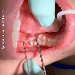
| “The gingival flap distal to an erupting molar, an operculum, can cause a lot of havoc.” |
| An operculum can lead either to pain caused by the occlusion of the opposing maxillary molar or by local inflammation/infection (pericoronitis) because of food debris and plaque accumulation between the tooth surface and the gingival flap. |
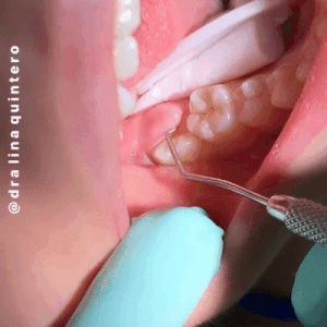
The painful and/or inflamed/infected operculum can be removed by operculectomy—the surgical excision or ablation of the operculum.
Laser therapy is an effective and noninvasive treatment option for Operculectomy. Lasers offer different wavelengths like 450nm/810nm or 980nm, out of which 450nm can be used in non-contact/contact mode whereas 810nm and 980nm are used in contact mode to perform operculectomy.
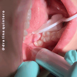
In this case, a wavelength of 450nm was used for the excision of inflamed pericoronal flap under topical anesthesia and crown of the tooth was exposed which further cleared the path of eruption.
Khan MA et al in 2017 used 980nm wavelength for operculectomy and suggested that due to many intraoperative and postoperative advantages, diode laser has become a preferred option for minor oral surgical procedures. Fornaini C et al. in 2016 also concluded that the 450 nm wavelength works very efficiently in the oral soft tissue surgical procedures, with faster healing and lesser thermal damage.
| Lasers offer certain advantages like the excellent hemostatic property which is of great value and allows the surgeon to work with better visibility, minimum patient discomfort, reduced post-operative pain, less chair side time, reduced healing time, reduction in edema and post-operative bleeding. |
| Extra Reading – Dental Lasers Overview (For The Motivated) By Dr. Martin Jablow, New Jersey. |
| Laser is an acronym that stands for Light Amplification by Stimulated Emission of Radiation. Light is measured in wavelengths. Dental laser wavelengths are Diodes 830-1,064nm, Nd:YAG 1,064nm, Erbium 2,790-2,940nm, and CO2 9.3-10.6nm. Every wavelength has a specific thermal output and specific tissue interaction that is always predictable. The different wavelengths for dental lasers perform different procedures. The most common dental lasers go by acronyms that are associated with how the laser light is produced. They include Diode, Nd:YAG, Er:YAG, or CO2. The dental laser manufacturers give the lasers names, but in many cases the wavelength of the laser is identical to another. It then becomes about the customization of the hardware and the number of parameters a dentist wants to control. Nd:YAG, Diode lasers, and CO2 are primarily used on soft tissue. These lasers provide good coagulation. The laser energy of the Diode and Nd:YAG can penetrate a few millimeters into the tissue. A CO2 laser penetrates less than a millimeter and can produce excellent coagulation along with a very precise cut. Erbium wavelength lasers can cut tooth, bone, and soft tissue. Erbium lasers have an affinity for water. Erbium lasers are not good at providing hemostasis but are kind to tissue, as they only penetrate microns into tissue. Periodontal procedures such as the laser-assisted new attachment procedure (LANAP) allow for minimally invasive surgery as an alternative to conventional flap surgery. The laser is placed into the periodontal sulcus and removes the diseased epithelium. Scaling is performed to remove calcified plaque and calculus from the root surfaces. The Nd:YAG produces a bactericidal effect in the pocket. Some individuals have had regeneration of cementum, bone, and periodontal ligament while reducing pocket depth. Nd:YAG, Diode, CO2, or Er:YAG lasers can also be used for the destruction of aphthous ulcers. A laser can transform a sore that hurts into a sore that does not hurt, with more rapid healing. Advantages of soft-tissue lasers may include minimal postoperative pain, minimally invasive surgeries, and in many cases, the elimination of sutures. Second-stage implant surgery can be safely accomplished with Diode, Erbium, and CO2 lasers. The KaVo DIAGNOdent uses a laser to help identify caries. Diagnostic lasers can be used to detect smooth surface, interproximal, and pit and fissure caries. These lasers operate at a wavelength of 655nm and are red in color. At this wavelength healthy enamel and dentin have little or no fluorescence. The laser light causes the carious areas of the tooth to fluoresce. The fluorescence is then measured, and the determination of caries can be made. Laser caries detection offers greater sensitivity than conventional visual and tactile methods. Early diagnosis of carious lesions makes minimally invasive dentistry possible. Erbium-based lasers (Er:YAG and Er,Cr:YSGG) can do both hard- and soft-tissue procedures. Many general dentists are looking to lasers to prepare teeth for restorations. Erbium lasers are effective for caries removal and cavity preparation. Many procedures can be done with an Erbium laser without anesthesia, and these lasers make treating pediatric patients easier. Erbium lasers do not cut teeth as fast as a high-speed handpiece, but they are much more conservative when preparing teeth. Erbium lasers can be used to remove composites but not amalgam restorations. Soft-tissue procedures with an Erbium laser can produce more bleeding compared to soft-tissue-only lasers. |
Today’s Morning Huddle was brought to you by Dr. Alina Quintero.
Instagram – @dralinaquintero