The ultrastructure of epidermal surface and stomata in subgenus Hippochaete (Equisetum, Equisetaceae)


The ultrastructure of epidermal surface of stem internods of horsetails of subgenus Hippochaete (Equisetum, Equisetaceae)

by Feoktistov D. S., Gureyeva I. L. (2016)

Dmitriy Feoktistov, Tomsk State University, Russia

Ii_Gureyeva
I.I. Gureyeva, Tomsk State University, Russia

===

in Turczaninowia DOI 10.14258/turczaninowia.19.3.2 –

https://www.researchgate.net/publication/313382656_The_ultrastructure_of_epidermal_surface_of_stem_internods_of_horsetails_of_subgenus_Hippochaete_Equisetum_Equisetaceae

Abstract
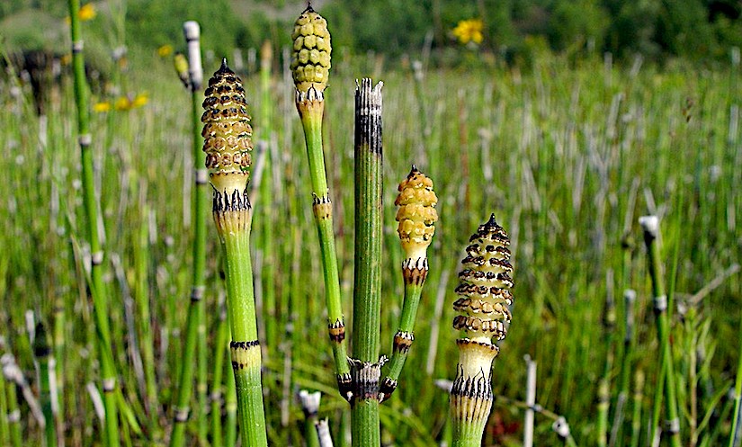
Ultrastructure of epidermal surface of stem internodes of 4 species and 2 interspecific hybrids of subgenus Hippochaete (Milde) Baker (Equisetum L., Equisetaceae) were studied using scanning electron microscopy: Equisetum hyemale L., E. ramosissimum Desf., E. scirpoides Michx., E. variegatum Schleich. ex F. Weber et D. Mohr, E. × moorei Newman, E. × trachyodon (A. Braun) W. D. J. Koch.
Epidermal ornamentation of stem internodes in subgenus Hippochaete is fundamentally different from that in subgenus Equisetum. The most visible elements of the surface, specific for the members of subgenus Hippochaete, are the stomatal passages, surrounded by bulky thickenings formed from the fused knobs.
Stomatal passages arranged allow the flanks of the furrows may be round, elliptical or almost rectangular; thickenings can be oblong, circular or horseshoe-shaped. The distinctly prominent sculptural elements of the furrow surface are knobs that may be discrete, arranged in chains or fused into irregular bars.
The relatively large sculptural elements – tubercles – are situated on the ridges and mostly arranged in two longitudinal rows. The wide ridges have additional fine ribbing. In some species, the hemispherical verrucae cover on the surface of the furrows and ridges.
The combination and arrangement of sculptural elements are more or less specific to the species. Features characteristic to the parental species are combined in ornamentation of the epidermal surface of stem internodes of hybrids. The detail descriptions of the sculpture of the epidermal surface of stem internodes are given for all studied species and hybrids.

The ultrastructure of epidermal surface and stomata of subgenus Equisetum


The ultrastructure of epidermal surface of stem and branch internods and spores of horsetails of subgenus Equisetum (Equisetum L., Equisetaceae)

by Feoktistov D. S., Gureyeva I. L. (2016)

Dmitriy Feoktistov, Tomsk State University, Russia

Ii_Gureyeva
I.I. Gureyeva, Tomsk State University, Russia

===

in Turczaninowia DOI 10.14258/turczaninowia.19.1.6 –

https://www.researchgate.net/publication/299459156_The_ultrastructure_of_epidermal_surface_of_stem_and_branch_internods_and_spores_of_horsetails_of_subgenus_Equisetum_Equisetum_L_Equisetaceae

Abstract
Ultrastructure of epidermal surface of stems and branches internodes of 5 species and 3 interspecific hybrids were studied using scanning electron microscopy, as well as spore surface of 5 species of horsetail subgenus Equisetum (Equisetum, Equisetaceae): E. arvense L., E. fluviatile L., E. palustre L., E. pratense Ehrh, E. sylvaticum L., E. × mildeanum Rothm. (E. pratense x E. sylvaticum), E. × sergijevskianum C. N. Page et Gureeva (E. pratense × E. palustre), E. × lofotense Lubienski (E. arvense × E. sylvaticum).
Sculptural elements from silica differ in shape, size and location on the ridges and furrows of stem and branches internodes. There are: the thin longitudinal sometimes anastomosing thread-like structures (ribs), cylindrical, rounded on the tip, or conical mamillae, tubercles and spines.
The thin longitudinal ribs, tubercles and spines are situated on the ridges of stem and branches; separate mamillae and groups of fused mamillae occupy the furrows. The fine surface sculpturing consists of hemispherical, globose or slightly elongate pilulae covering mamillae, surface in furrows, and especially stomata.
Equisetum palustre has specific club-shaped rods arranged into regular ranks along either side of the stomatal slit. A detailed description of silica sculpture, shape, disposition and ornamentation of stomata area are presented for all studied species and hybrids.
Such features as the presence, shape and arrangement of sculpture elements, ornamentation, shape and arrangement of stomata can be used in the taxonomy and phylogenetics of horsetails. Spores of the members of subgenus Equisetum are spherical in shape. They have not specific ornamentation, their features may not be used in systematics of horsetails.

Stomata in Equisetum (in German)

Photo credit: Google

Spore cones: Equisetum hyemale. ~ By Donald Cameron.

Screen Shot 2017-09-14 at 10.54.34


Untersuchung der Spaltöffnungsapparaten von Equisetum

by Kedves M. (1958)

Universität Szeged, Hungary

in Acta Biol. Szeged 4: 149-155. –

http://acta.bibl.u-szeged.hu/21464/1/biologica_004_149-155.pdf

Screen Shot 2017-09-14 at 10.59.22

Einleitung

Die von anderen Typen der Pflanzenwelt abweichende Form der kompliziert aufgebauten Stomen der Equiseten hat RIEBNER (1926) — nach der Überprüfung der Arbeiten von vorigen Autoren — festgestellt. Er hat sich eingehend mit den physiologischen Fragen beschäftigt, und die systematischen Beziehungen hat er unbeachtet gelassen. Aus diesem Gesichtspunkt ist die zusammenfassende Arbeit von MII.DE (1867) wichtig, aber seine, über die Spaltöffnungsapparaten mitgeteilten Zeichnungen sind sehr abweichend von denen, die in RIEBNER’s Arbeit mitgeteilt sind. Deshalb war ich bestrebt die Frage am Grunde genauer morphologischer Untersuchung aus systematischem und — von den bisherigen Auktoren abweichend — aus entwicklungsgeschichtlichem Gesichtspunkt zu lösen.

Screen Shot 2017-09-14 at 10.57.13

Zusammenfassung

Die Spaltöffnungsapparate von E. xylochaetum Mett., E. pyramidale Goldm. und E. Martii zeigen einen übergänglichen Charakter zwischen den phaneropor und cryptopor Stomen. Der Aufbau und die Verzweigung dieser Arten steht auch zwischen den zwei Typen, deshalb ist es begründet die Equiseten — statt dem bisher in zwei gegliedertem System — in drei Sektionen zu teilen.

Stomata in Equisetinae and Lycopodinae

screen-shot-2017-02-21-at-18_fotor

Über Bau und Funktion der Spaltöffnungsapparate bei den Equisetinae und Lycopodinae

by Riebner F. (1926)

Fritz Riebner – Universität Berlin

in Planta 1: 260-300 – doi:10.1007/BF02038171

http://link.springer.com/chapter/10.1007%2F978-3-642-49824-4_7

screen-shot-2017-02-21-at-19-04-35

Zusammenfassung

Die Equisetinen haben durch ihre isolierte Stellung im Pflanzenreiche schon frühzeitig das Augenmerk der Forscher auf sich gelenkt, wobei auch ihre Anatomie mehrfach studiert wurde.

Dabei fiel besonders der Spaltöffnungsapparat dieser Pflanzengruppe auf, dessen Bau verschiedene Eigentümlichkeiten zeigte, die allen übrigen Pflanzen fehlen.

screen-shot-2017-02-21-at-19-07-47

Alle bisherigen Arbeiten darüber sind aber wenig eingehend; die Beschreibung der Stomata ist manchmal recht ungenau und teilweise sogar offensichtlich falsch. Die Autoren begnügen sich ferner mit einer rein deskriptiven Behandlung des Objektes, und es ist noch kaum der Versuch gemacht worden, Aufschlüsse über die Bewegungsmechanik dieser Spaltöffnungen zu erhalten.

screen-shot-2017-02-21-at-19-11-03

Diese klar zu legen war das Ziel meiner Arbeit. Um dafür die notwendigen Grundlagen zu gewinnen, nahm ich zunächst eine möglichst eingehende Untersuchung des anatomischen Baues der Spaltöffnungsapparate einiger Equisetum-Arten vor, um dann auf Grund von Beobachtungen der Schließbewegung und von Messungen möglichst Klarheit über die Funktion zu schaffen.

Um endlich den Vergleich der EquisetumSpaltöffnungen mit denen aller übrigen Pteridophyten zu ermöglichen, wurden noch einige bisher wenig untersuchte Gruppen derselben bearbeitet.

Stomata in Equisetum scirpoides


Origin and development of tissues in Equisetum scirpoides

by Johnson M. A. (1933)

Marion A. Johnson

Hull Botanical Laboratory

in Bot. Gaz. 94 : 469-494. –

https://www.jstor.org/stable/2471233?seq=1#page_scan_tab_contents

Abstract

1. The first periclinal division in each segment from the apical cell of the stem separates pith and “primary cortex,” which in turn gives rise to stele and cortex.
2. The pericycle, endodermis, and one or two layers of the inner cortex of the stem are stelar in origin.
3. Evidence is presented for regarding the so-called inner endodermis of the root as pericycle.
4. The endodermis and pericycle of the root are of cortical origin.
5. Dormant branches occur as in other species of Equisetum. They are exogenous in origin. 6. The first root developed by the branch is endogenous.
7. The stelar parenchyma in the root is much reduced.
8. Hydathodes of the water stomata type occur on the upper surface of the leaves.
9. Rare but well-developed tyloses are found in the carinal canals.
10. Reduction in xylem is extreme, the supranodal wood being frequently absent above the leaf traces, forming a gap in the xylem.
11. The gaps in the xylem of the cone are correlated with the size of the stele and failure of the fundamental tissue to develop.

Etude ontogénétique et ultrastructurale des stomates de l’Equisetum arvense L.


Contribution à l’étude du transit du silicium chez l’Equisetum arvense L. I. Les stomates : Etude ontogénétique et ultrastructurale

par Laroche J., Robert D., Le Coq C., Guervin C. (1976)

in Rev. gén. Bot. 1976, 83, 331-365.

screen-shot-2017-01-10-at-18_fotor

screen-shot-2017-01-10-at-18_fotor-b

screen-shot-2017-01-10-at-18_fotor-c

screen-shot-2017-01-10-at-18_fotor-d


Stomata in Equisetum


The stomatal apparatus of Equisetum 

by Hauke R. L. (1957)

Richard L. Hauke, University of California, Berkeley

in Bulletin of the Torrey Botanical Club 84:178-181. – DOI: 10.2307/2482890 – 

https://www.jstor.org/stable/2482890?seq=1#page_scan_tab_contents

Abstract

1. A long-standing discrepancy in the literature pertaining to the location of the radiating bands in the stomatal apparatus of the genus Equisetum was discussed, with the citation of six authors who stated that they were part of the subsidiary cell wall, and another six authors who stated that they were part of the guard cell wall.
2. Macerations were made of desilicified and cleared material of twelve species of Equisetum, which revealed that these bands are always located on the subsidiary cell wall, thus confirming Sanio’s original report.


Development of stomata in Isoetes, Lycopodium, Selaginella and Equisetum

Photo credit: Google

Development of stomata in some fern allies

by Pant D. D., Mehra B. (1964)

Divya Darshan Pant, Bharati Mehra

in Proceed. Nat. Inst. Sci. India  Vol. 30, B, No. 2: 92-98

http://www.dli.gov.in/rawdataupload/upload/insa/INSA_1/20005b76_92.pdf

Abstract

screen-shot-2016-12-08-at-00-02-01

screen-shot-2016-12-08-at-00-30-09

screen-shot-2016-12-08-at-00-35-30

screen-shot-2017-02-21-at-19_fotor

screen-shot-2017-02-21-at-19_fotor

Development of stomata in Equisetum


Development of stomata in Equisetum

by Pant D. D., Kidwai P. F. (1968)

in Ann. of Bot. 32 (127): 601. –

http://aob.oxfordjournals.org/content/32/3/601

Abstract

Hitherto published accounts of the development of stomata in Equisetum are conflicting about the sequence of divisions and about the formation of a sub-stomatal cell from a meristemoid.

The present study of the development of stomata in a species identified as E. ramosissimum subspecies ramosissimum supports the observations of Strasburger (1867) and Pant and Mehra (1964) on the basis of sections cut in various planes through internodes of the plant.