The gametophytic stage of this species is relatively distinct in the NW Atlantic flora being pyramidal in outline and feathery in appearance (Image A). This stage is typically found entangled with other species via its distinctive hooks (Image B). In surface view the smaller hyaline gland cells are obvious among the regular vegetative cells (Image C). Tips of indeterminate branches are uniseriate with a rounded apical cell (Image D) whereas tips are typically acute for lateral branchlets (Image E). The sporophyte (Trailiella phase) is common, but more cryptic in appearance relative to many other filamentous species (Image F), but stands out for its prolific production of distinctive gland cells at the nodes between adjacent barrel-shaped cells and tendency to produce branches at 90o angles (Image G). Although commonly found unattached in the drift, the sporophyte also grows attached to various hard substrata and other algae. When this occurs a prostrate filament sends up erect filaments at 90o angles and is attached by a complex of rhizoids (branched uni or multicellular(?); Image H). Cruciate tetrasporangia are produced in some regions of our flora, forming singly or in short series of 2-5 on the sporophytic filaments (Image I).
Widely distributed in both stages in New England waters of the NW Atlantic, the sporophyte is also common in the Maritime provinces, while the gametophyte has become more prevalent in the latter region over the past decade. An introduced species, we have genetically verified records of the gametophyte from RI to the Bay of Fundy, NB, and of the sporophyte extending to southern and western NL. There is a slight chance of confusion of the sporophyte stage with Spermothamnion repens (Dillwyn) Magnus and Spermothamnion cf. roseolum (C.Agardh) Pringsheim, but these species have wider main axes, commonly with opposite branching, and lack the characteristic glands cells of Bonnemaisonia hamifera.
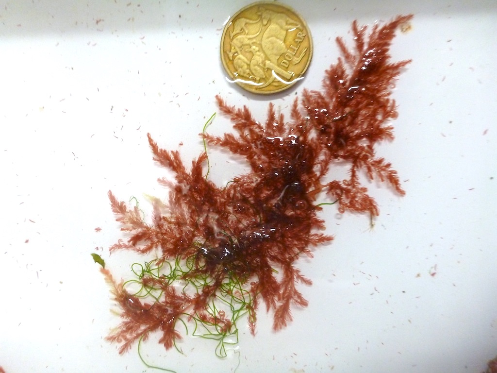 Image A. Gametophyte stage tangled in algae in the low intertidal from Maces Bay, Bay of Fundy, NB (GWS034091).
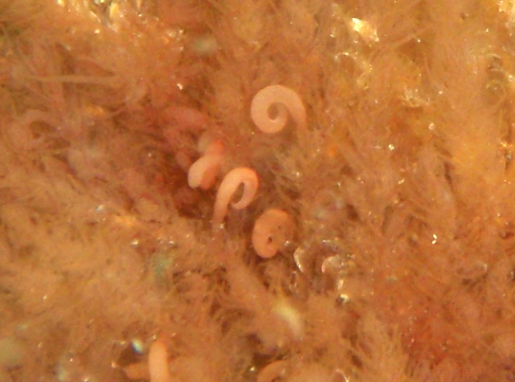 Image B. Close-up view of the hooks formed on the gametophytic stage (specimen from Folly Cove, Gloucester, MA).
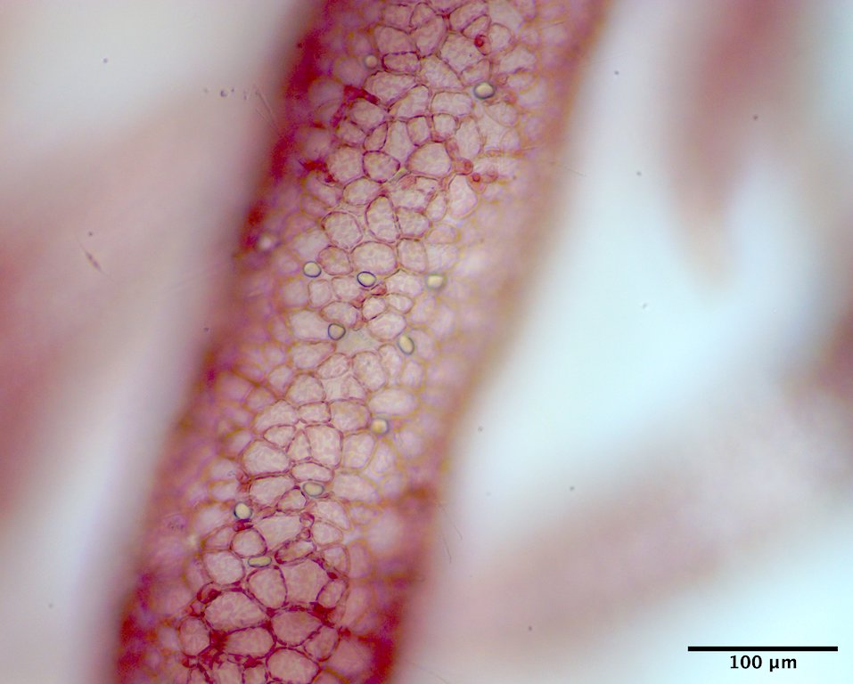
Image C. Surface view reveals gland cells among vegetative cells on the gametophyte surface (drift, Maces Bay, Lepreau, Bay of Fundy, NB; EGWS001126).
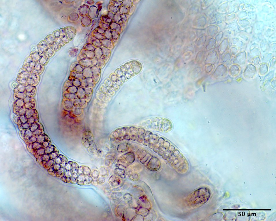
Image D. Indeterminate branch tips, uniseriate with rounded apical cells (EGWS001126).
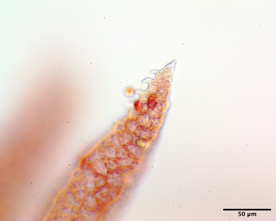
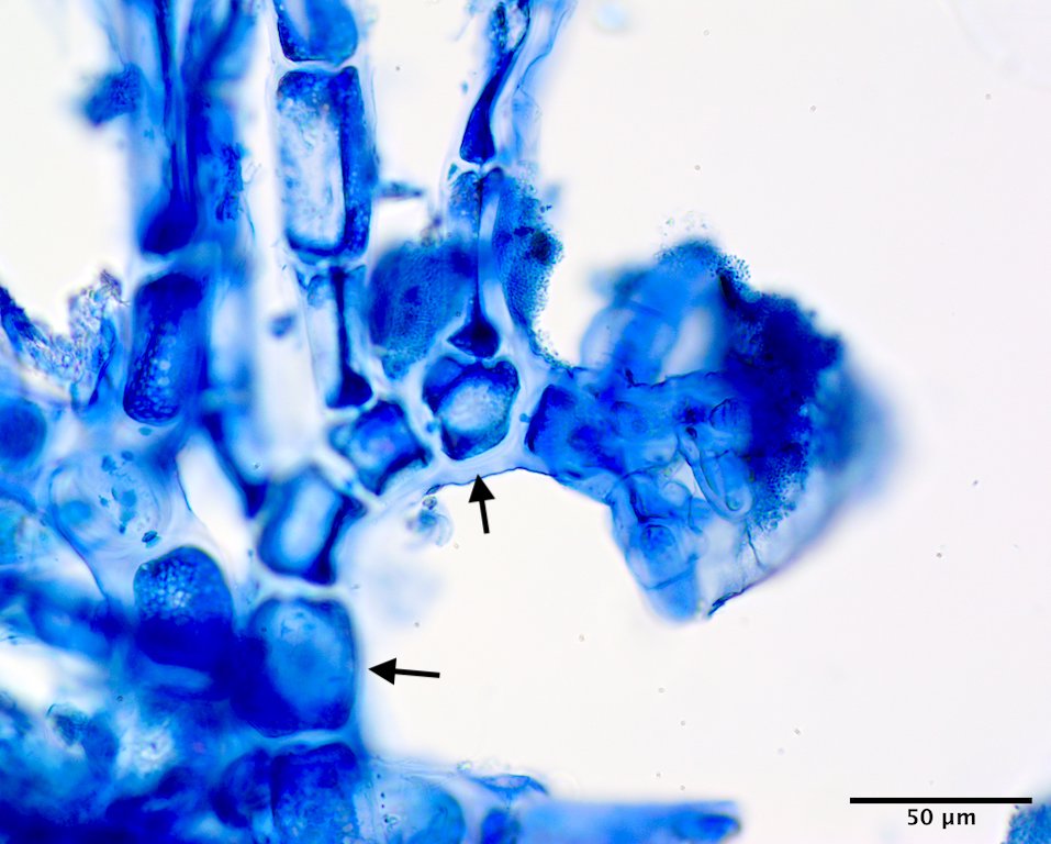
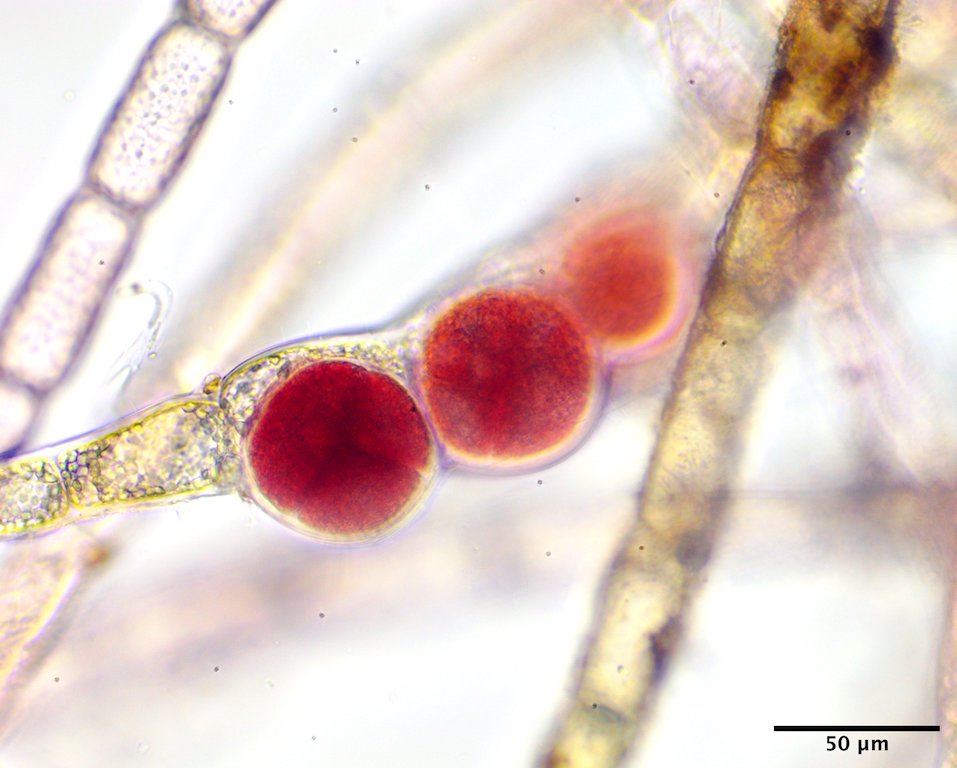
Image E. Lateral branchlet tips with acute apical cells (EGWS001126). Image H. Trailiella phase growing subtidal (20 m) on Laminaria solidungula J.Agardh; a prostrate filament (arrows) anchored by a rhizoidal base and producing erect filaments (South side of bay mouth, Bonne Bay, NL; GWS007237; rehydrated from press; aniline blue stained). Image I. Tetrasporangia in various stages of development (leftmost mature) (drift, Beach to east of Parlee Park Beach, NB; GWS044435).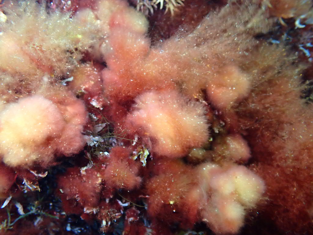 Image F. Sporophyte (Trailliella phase) subtidal (3 m) growing on algae at Birchy Head, NS (EGWS000551).
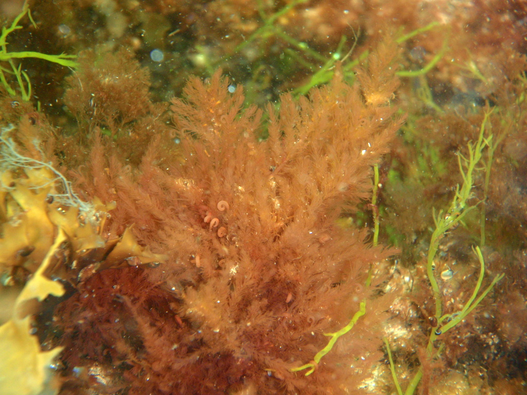
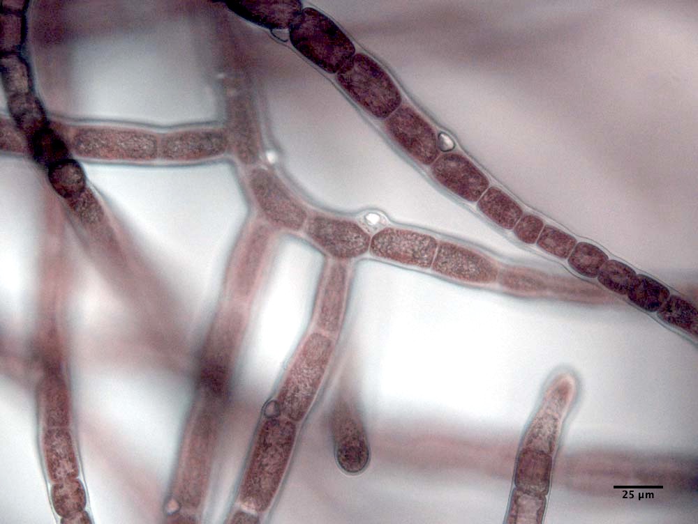 Image G. Trailiella phase with right angle branching and gland cells evident (subtidal (6 m), on Polysiphonia; The Rock, Bras d’Or, Cape Breton; GWSC049, culture).