Article

Morphoanatomical, Histochemical, and Essential Oil Composition of the Plectranthus ornatus Codd. (Lamiaceae)

by
Luiz Renan Ramos da Silva
1,
Zelina Ataíde Correia
1,
Ely Simone Cajueiro Gurgel
1,
Olívia Ribeiro
1,
Sebastião Gomes Silva
2,
Oberdan Oliveira Ferreira
2,
Eloisa Helena de Aguiar Andrade
1,2 and
Mozaniel Santana de Oliveira
1,2,*
1
Postgraduate Program in Biological Biological Sciences–Tropical Botany, Universidade Federal Rural da Amazônia and Museu Paraense Emílio Goeldi, Av. Perimetral, 1901, Terra Firme, Belém 66075-900, Brazil
2
Adolpho Ducke Laboratory—Botany Coordination, Museu Paraense Emílio Goeldi, Av. Perimetral, 1901, Terra Firme, Belém 66077-830, Brazil
*
Author to whom correspondence should be addressed.
Molecules 2023, 28(18), 6482; https://doi.org/10.3390/molecules28186482
Submission received: 3 August 2023 / Revised: 31 August 2023 / Accepted: 2 September 2023 / Published: 7 September 2023
(This article belongs to the Special Issue Plant Secondary Metabolites in Medicinal and Aromatic Plants)

Abstract

:
Plectranthus ornatus is a medicinal and aromatic plant used in traditional and alternative medicine. In this study, leaves of P. ornatus were collected in two cities of the state of Pará, “Quatipuru” and “Barcarena”, and were used with the objective of analyzing, through morphoanatomical data and histochemical and phytochemical studies of essential oil, the samples present structural differences and differences in their chemical composition. Anatomical and histochemical analyses were performed by transverse, using longitudinal sections of 8 μm to 10 μm to perform epidermal dissociation, diaphonization, and tests to identify classes of secondary metabolites. The essential oils were isolated by hydrodistillation, and the identification of the chemical composition was performed by gas chromatography coupled with mass spectrometry. The anatomical study shows that there is no difference between specimens collected in different locations, and stellate trichomes were identified. The histochemical study detected total lipids and acids, terpenes, polysaccharides, phenolic compounds, tannins, alkaloids, and calcium oxalate. The low essential oil yield may be related to the low density of secretory cells (glandular trichomes), the unidentified compounds in the highest concentration in the essential oil were in relation to the chemical composition of the essential oils, and the major compounds were α-pinene, sabinene, (E)-caryophyllene, caryophyllene oxide, and oct-1-en-3-ol. The results provide new information about the anatomy and histochemistry of P. ornatus.

Graphical Abstract

1. Introduction

The most famous plants used in traditional medicine are from the Plectranthus genus, belonging to the Lamiaceae family, including around 200 genus and 3200 vegetable species, and the subfamily Nepetoideae, which incorporates plants commonly known as “boldo” [1]. The name Plectranthus is derived from the Greek words plektron (spur) and anthos (flower), describing a stimulus that the flowers have in their support [2]. Many Plectranthus species are rich in essential oils, compounds produced in the secondary metabolism of plants. These compounds have demonstrated several applications in the most diverse industrial segments, such as cosmetics, pharmacology, and the food industry [3,4,5], because the complex mixture of the molecules that form essential oils can present potential biological activities. For example, Plectranthus ornatus is used in herbal medicine for its potential health benefits [6].
Essential oils (EOs) are formed by complex mixtures of volatile substances. These oils are biosynthesized in the secondary metabolism of plants, and several classes of compounds can be identified in essential oils such as monoterpenes, sesquiterpenes, and phenylpropanoids [7,8,9,10,11]. These complex mixtures is strongly volatile according to abiotic environmental factors. They are colorless to slightly yellow and capable of dissolving in other oils, fats, wax, and ethanol [12]. Their functions include protection, internal and external dispersion against infectious agents on the plant, and attraction of pollinators [13].
Species of the genus Plectranthus are characterized by rapid growth and can tolerate a lack of natural resources such as sunlight and rain. In addition, these special species are easy to adapt to the most diverse biomes that can be incorporated, both in Brazil and in other countries [2]. Despise their origin elsewhere, they are notoriously mentioned in Brazil, especially the genus Plectranthus. Their diverse use in medicine is reported in ethnobotanic data, and their leaves treat headaches, stomachaches, dermatitis, burns, and allergies to insect bites and act as sanitizers [14].
Moreover, it is difficult to identify the genus Plectranthus due to morphological similarities among the species, which contributes to obstacles in defining criteria for taxonomic identification of the genus [15]. Another problem for taxonomic identification is the common names, because in Brazil, for example, there are five different species of the genus Plectranthus known by the same common name, “boldo.” Therefore, anatomical and histochemical studies in addition to the chemical composition of essential oils can also be used as a marker of the species or genus Plectranthus [16].
It is known that the species P. ornatus (boldo miúdo, boldo-chinês, falso boldo) disseminates easily as cultivated or spontaneously, is a remarkable ornament, therapeutic through its secondary metabolites (where diterpenes are the main class of substance), and its morphological aspects may vary according to the habitat where it is placed [10]. However, few studies refer to the anatomical and histochemical approaches of the species P. ornatus. In addition, the struggle with properly identifying the species in this genus and their relevance due to medicinal properties might contribute to future surveys from presented Morph anatomical and histochemical data. These plant species are rich in secondary metabolites with applications in various industrial segments [17]; therefore, it is necessary to know the classes of these metabolites and their secretory cells. In this sense, the present work had as its objective to analyze if two samples of P. ornatus, collected in different cities of the state of Para-Brazil, have anatomical differences, histochemical differences, and differences in the chemical composition of the essential oils.

2. Results and Discussions

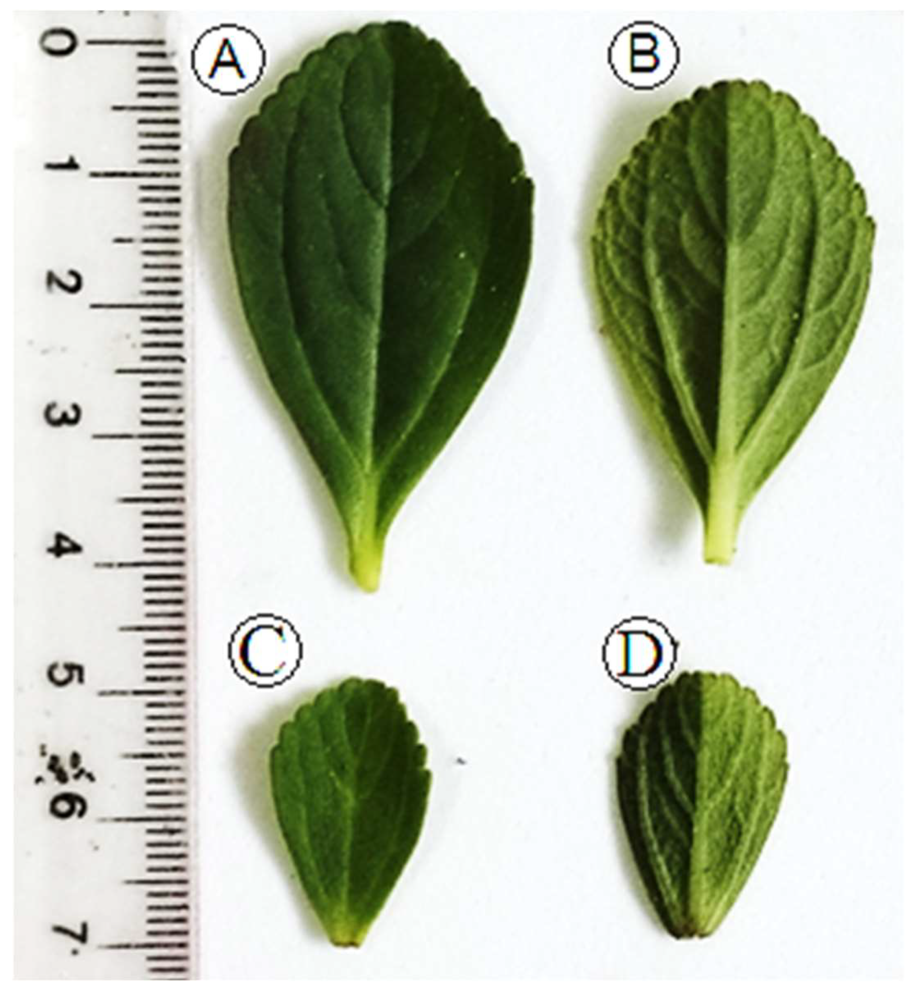
2.1. Morphological Aspects

Morphologically, P. ornatus presents with vegetative characteristics similar to other species of the genus Plectranthus (Lamiaceae), such as P. barbatus, P. neochilus [14], P. amboinicus [18], P. verticillatus [19], and P. neochilus [20]. This can also be one of the factors that can hinder the correct taxonomic identification of the species of the genus Plectranthus. In the present work, specimens A and B present the same morphological characteristics. The differences were only in the size of the leaves, as can be seen in Figure 1. The leaves presented morphological characteristics of opposite arrangement. They are simple, with an elliptical oval shape, have a dentate margin in the upper half, have small petioles, and are peninerveous. Both the adaxial and abaxial parts have a pilose and succulent surface, that is, they store water and are a kind of mucilage deposit, which can protect the plant from dehydration [21]. This species is characterized by a strong aroma, which can characterize a potential for production and secretion of EOs [22]. Those morphological aspects are confirmed by reported data from the Global Biodiversity Information Facility (GBIF) and Reflora—Espécies da Flora do Brasil about P. ornatus Schltr.

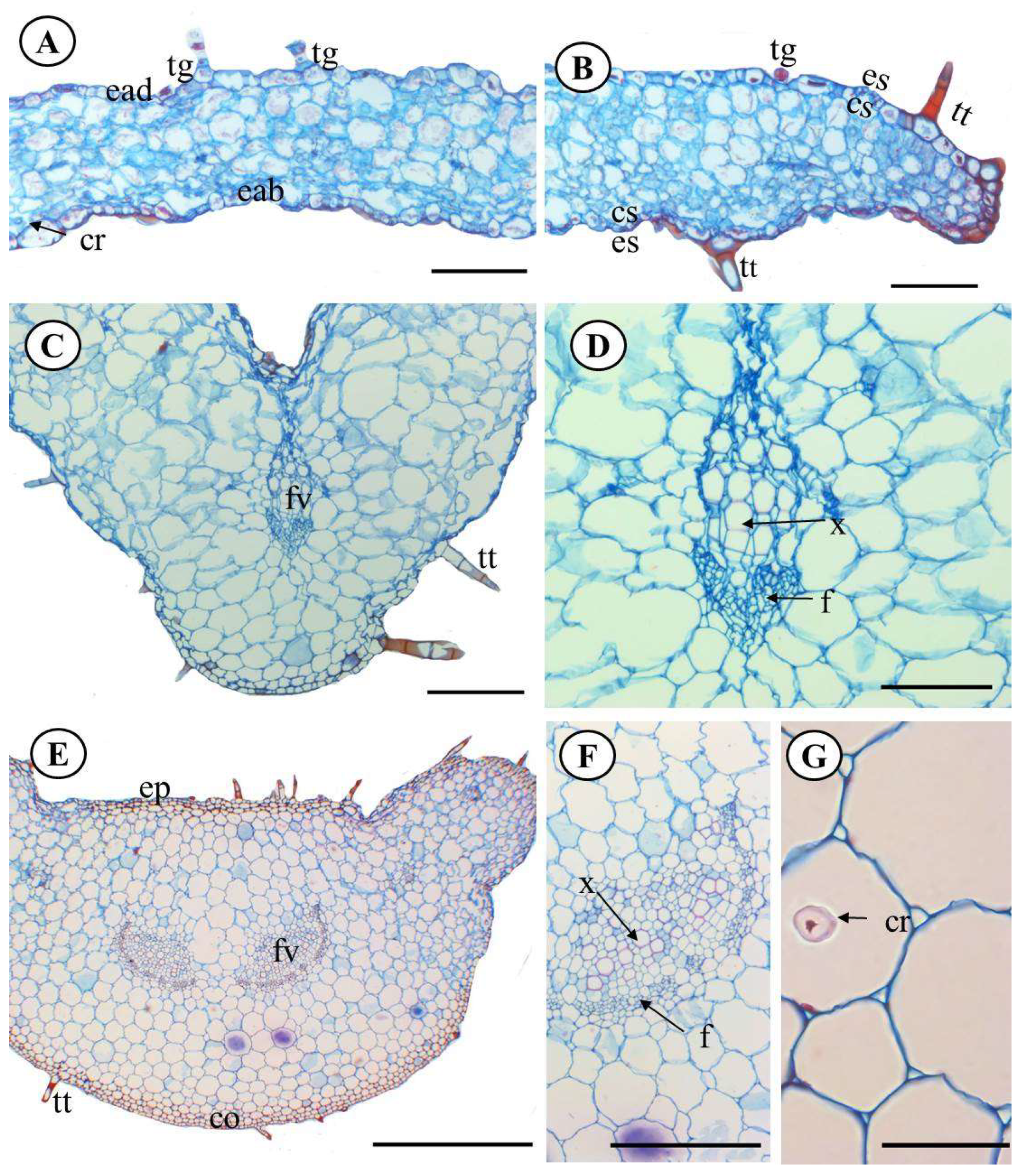
2.2. Anatomical Characterization
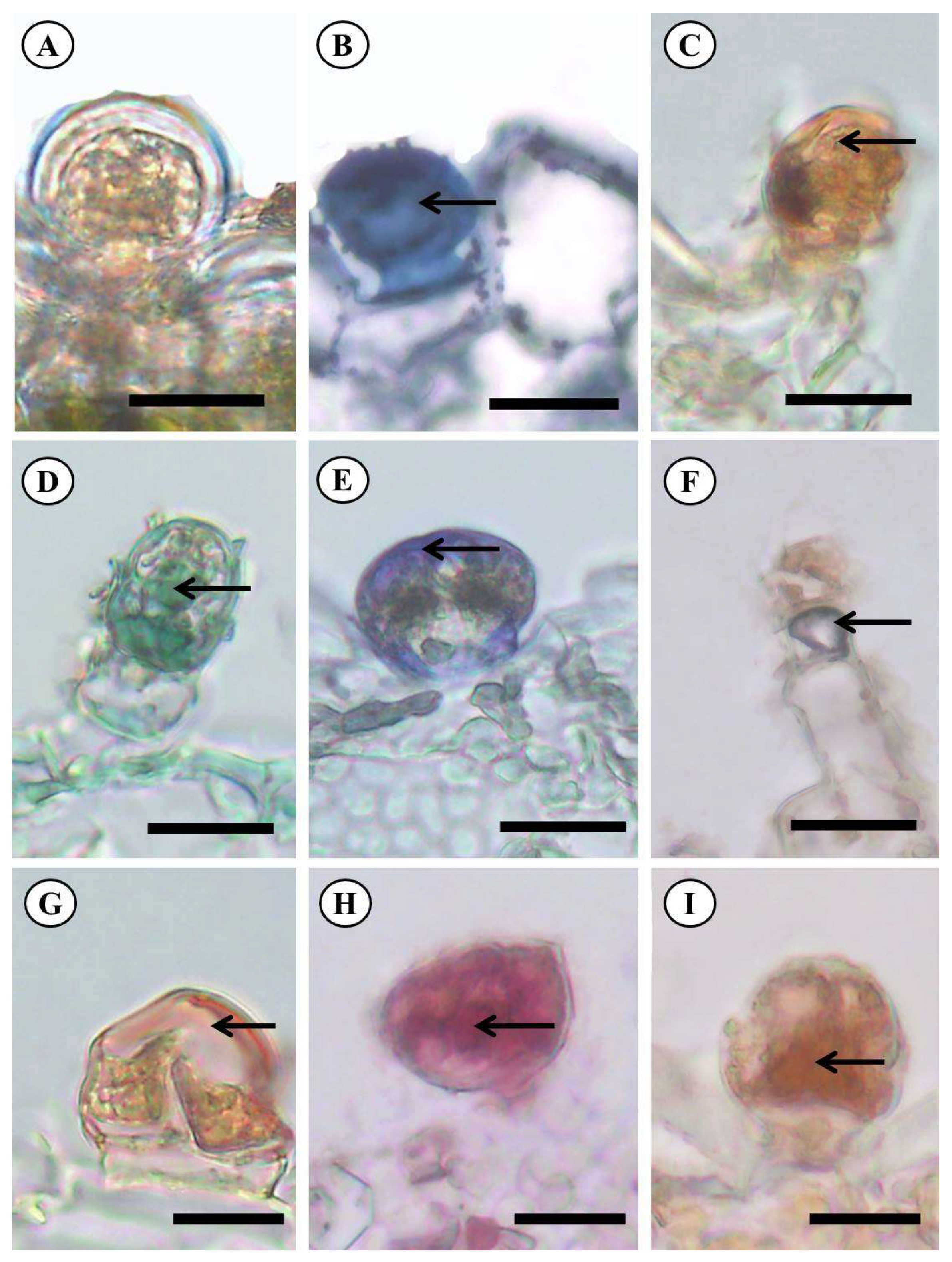
The anatomical characterization of species P. ornatus exhibited that the leaf has in both adaxial and abaxial epidermal sides the presence of epidermal cells and sinuous anticlinal walls (Figure 2A,B). The leaf blade was amphistomatous, and the stomata were diacytic (Figure 3B). Mauro et al. [23], analyzed the anatomical structures of P. ornatus and characterized the stomata in a paradermal view, belonging to the anomocytic (irregular-celled) type and presenting reniform guard cells, immersed in epidermal cells with a sinuous contour; however, our results indicate that this classification may not be correct, as the subsidiary cells are organized in such a way that they form a section perpendicular to the stomatal cleft.
Distinct trichomes were found on the two sides of the leaf, being classified as pluricellular uniseriate tectors and glandular as two descriptive characteristics for this species (Figure 2C–E) and representing the main secreting structure found on the leaf of P. ornatus. Starred trichomes were also observed (Figure 2F). The trichomes are notable epidermal appendixes for defining the identification within the genus Plectranthus [14] for taxonomic and medicinal/pharmacological criteria. As a counterpoint, starred trichomes have not been found in the research of Mauro et al. [23], but these characteristics are proven for the species P. ornatus as identified by Ribeiro et al. [24]. In general, the anatomical analyses showed no difference between the specimens studied, as can be seen in Supplementary Material S1.
The cross-sectional area showed that the median leaf region is constituted by uniseriate epidermis and tabular and juxtaposed cells, covered by a smooth and slim cuticle, pluricellular non-glandular trichomes (two to three cells), uniseriate, in a tapered shape and glandular, showing pedicel trichomes, and also sessile glandular trichomes (Figure 3A,B). This morphological trichomes variation on the studied species has been predicted by previous studies [23,24].
A homogeneous mesophile was noted, with no distinction between the palisade and spongy parenchyma and prismatic crystals of calcium oxalate in the idioblasts (Figure 3A). The boundary region was a rounded shape and had compacted cells (Figure 3B).
The cross-sectional area showed that the midrib has a convex format. The epidermis is uniseriate, tabular, and juxtaposed and has a smooth cuticle (Figure 3C). The midrib cortex was formed by 2–3 layers of angular collenchyma next to the epidermis. That is explained by the irregular distance of the cell wall from the sustenance system because it is a mechanical resistance from the vegetative organs still growing [25].
The parenchymal tissue was formed by polygonal, heterodimensional, thin-walled, and slightly sinuous cells. In the central area of parenchymal 1, a collateral vascular bundle was detected (Figure 3D). Those perceptions have also been reported by Mauro et al. (2008) and Ribeiro (2017), as well as for other species belonging to the family, like species Leonurus sibiricus L [26].
The petiole on the cross-sectional area had common characteristics with Peumus boldo, P. neochilus, P. verticillatus, and P. amboinicus [19,23,24,26], such as convex format and projections on the sides. The epidermis was uniseriate and composed of rectangular diminished cells, where both glandular and non-glandular trichomes were present (Figure 3E).
The cells were polygonal, heterodimensional, and thin walled with slight sinuosity (Figure 3E). The vascular bundles were collateral, sclerenchyma-closed, surrounding the phloem, and two secondary bundles were also present in the sideways (Figure 3F). Crystals of calcium oxalate in the idioblasts were also noted (Figure 3G).
When the xylem and phloem are placed on opposite sides, as labeled in the anatomy of the species, the classification is “collateral” as predicted by the guidelines of morphological and anatomical classification of [27].
The presence of calcium oxalate, which was more concentrated in the petiole, reflects an adaptation in plants derived from differences in external factors, such as herbivory differences and formation of vegetative structures, and turns them into toxic ones [28], as observed in Plectranthus neochilus Schltr and reported by Duarte et al. [26].

2.3. Histochemistry

Histochemical tests suggested in this survey that the margin and petiole of P. ornatus reacted for distinct classes of secondary metabolites in glandular trichomes. According to Kalicharan et al. [29], global characterization detection usually happens in the nucleus of glandular specialized cells in trichomes, and the reagents react more evidently when metabolites are identified on glandular secretions. This is derived from the ability of glandular trichomes to store and secrete an amount of secondary metabolites [30]. The results of histochemical analysis are reported in Table 1, and the reactions are expressed in (Figure 4A–I).
The trichome secretions of Plectranthus ornatus reacted (positive result) for total lipids and acidic lipids. However, there is a difference in lipidic composition, as evidenced by the Nadi reagent. This reaction stained purplish, indicating terpenes of different sizes (molecular masses), implicating a mixture of essential oil and resin [31]. The terpene identification is more effective when the reaction happens (positive result), as seen in this study and also in previous works with P. amboinicus, Mentha x villosa Huds [18,32]. Genus Ocimum terpenes can also be found in the stem [33].
Those oil compositions might influence the ecological relationship established between the plant and the environment [18]. Hence, environmental conditions/disturbances are also a strong influencing factor on the yield of other substances and chemical compounds such as EOs, because their production is related to the leaf’s protection against external factors such as herbivory factors. Their production also helps pollination and aids in herbal action on the human body when using traditional medicine, as it acts as an analgesic, an antibacterial, and an antioxidant [13,34].
The tests on the species showed the presence of phenolic compounds and tannin, which has also been found in other species belonging to the family Lamiaceae, along the leaves of Plectrathus amboinicus (Lour.) Spreng [18] and every vegetative part but the inflorescence of Ocimum basilicum and O. campechianum [32]. The work of Rocha et al. [33] confirmed the positive result for tannin in histochemical tests, potentializing the probable explanation of the resistance and binder power of this plant.
Phenolic compounds are widely found substances in vegetables according to Silva et al. [35]. This functional group highlights substances such as coumarins, lignin, quinones, tannins, and flavonoids [36]. Phenolic compounds in vegetables might act against herbivores and pathogens, promote cell reconstruction, and attract pollinizers [37]. They also have healing, antioxidant, anti-inflammatory, and antimicrobial action [38].
Glandular trichomes from the analyzed species were demonstrated to have alkaloids likewise observed in other literature [18,39], on the leaves of Plectrathus amboinicus (Lour.) Spreng, and by Alasbahi, and Melzig [40], on leaves of Plectranthus barbatus Andrews. The alkaloids are some of the most important nitrogenated compounds found in herbal vascular plants, providing herbivory protection, along with anesthetic, antitumoral, myorelaxant, and antimicrobial activities [41].
The positive result for periodic acid-Schiff (PAS) pointed to the presence of total polysaccharides in glandular trichomes of P. ornatus. The polysaccharides are stocking molecules of hydrophilic carbohydrates that function in cell wall formation, and by their chemical properties, they prevent damages caused by hydric stress [42,43]. On the other hand, the polysaccharides from exudates are produced as a defense mechanism against physical injuries and microbial attack [44]. Therefore, the most relevant secondary metabolites found in the secretion of glandular trichomes of P. ornatus have defensive action, which somehow helps with survival in the environment.

2.4. Chemical Composition of the Essential Oil

The essential oils of specimens (A) and (B), as shown in Table 2, did not yield properly (0.1 mL), but the chemical characterization was still performed. A total of 93.45% and 99.92%, respectively, of the 93 compounds were identified and are described in Table 3. The hydrocarbon sesquiterpenes play a role of 27.56% in the chemical profile of essential oil from specimen (A) collected in the city of Barcarena, Brazil, and 41.65% for (B) collected in the city of “Quatipuru”, Brazil. Sesquiterpenes were also detected in essential oil (EO) from other species of the genus Plectranthus. However, an amount of 63.1% of monoterpenes was identified in a sample of P. ornatus and was identified in a sample collected by other authors [45] in the state of Ceara, Brazil.
The major components in the essential oil of specimen (A) were (E)-Caryophyllene (12.84%), α-pinene (12.38%), and sabinene (8.72%). On the other hand, the EO from specimen (B) was found to have (E)-caryophyllene (29.61%), 1-octen-3-ol (13.92%), and terpinen-4-ol (5.66%). The results varied from what has been found in a sample of P. ornatus collected in Lisboa, Portugal, in which the predominant substances were oct-1-en-3-ol (13–31%), β-pinene (11–24%), α-pinene (11–19%), and β-caryophyllene (11%) [55].
Literature reports the presence of monoterpenes in essential oils from genus Plectranthus as observed in EO from P. amboinicus, which showed in its chemical profile the following substances: carvacrol (23.0%), camphor (22.2%), δ-3-carene (15.0%), γ-terpinene (8.4%), o-cymene (7.7%), and α-terpinene (4.8%) [56]. (E)-caryophyllene was also observed in other genus studies such as in P. grandis, which had a content of (E)-caryophyllene (38.25%) higher than that found in the present work. In addition, P. grandis EO exhibited sesquiterpene compounds such as α-copaene (13.23%) and germacrene (11.38%). In P. ornatus EO, the major chemical components were caryophyllene oxide (61.74%) and β-caryophyllene (10.65%) [57].
In the work of Passinho-Soares et al. [58], a sample of P. ornatus was collected from a parent plant (main plant) cultivated at the Pharmacy College of Federal University of Bahia (Universidade Federal da Bahia, UFBA), and the major components of its EO were α-terpinyl acetate and the monoterpenes (α-thujene, α-pinene, β-pinene, camphene, sabinene, and α-limonene). Another study also detected monoterpenes, such as α-thujene (4.40%), β-bourbonene (8.07%), and 1-octen-3-ol (11.85%), in the EO of P. ornatus [59].

3. Materials and Methods

3.1. Collecting Area for the Botanical Material

The botanical material was collected in two different cities in the state of Para, Brazil. Sample A was collected in a garden of a local producer in the city of Barcarena (state of Para) at the following geographical coordinate: latitude 01°30′21″ south and longitude 48°37′33″ west. Sample B was collected in the city of Qautipurú (state of Para) located at the geographical latitude 0°53′56″ south and longitude 47°0′40″ west. Samples of specimens of P. ornatus were identified and deposited in the Herbarium of Museu Paraense Emilio Goeldi, João Murça Pires, registered with number MG N° 245302.

3.2. Morphological Analysis

The morphological leaf parameters were analyzed under the eyes of taxonomic reviews [60] and specialized literature [61] about leaf morphology to characterize and analyze the vegetative parts such as apex, margin, foliage, midrib, phyllotaxy, and petiole using magnifying glasses. The aerial vegetative parts of the species were collected for anatomical and histochemical studies, developed in the Vegetable Anatomy Laboratory (Laboratório de Anatomia Vegetal), in the Botanic Coordination of Museu Paraense Emílio Goeldi Museum—LAVEG/COBOT/MPEG, and have undergone the following techniques.

3.3. Anatomical Analysis of the Leaf Blade by Light Microscopy (LM)

The samples (Figure 1) of the leaf blade were sectioned into the regions used in the anatomical study (margin, midrib, and petiole), fixed on FAE (formaldehyde, acetic acid, and ethanol 50%) [47] for 24 h for the structural study, in hydrophilic substances for histochemical tests, in NBF (neutral-buffered formalin) for 48 h for lipophilic substances [62] and stored in ethanol 70%. Figure 5 displays the image of the leaf sections of Plectranthus ornatus.
For laboratory analysis, the samples previously stored in ethanol 70% were submitted for dehydration in a growing butyric alcohol series (butyric tertiary alcohol) to include it in histological paraffin with DMSO (Paraplast®, ©Leica Biosystems, Teaneck, NJ, USA) [47]. Transversal and longitudinal sections from 8 μm to 10 μm were obtained with the aid of a Rotary Microtome (model Leica® RM 2245, Leica® Biosystems, Heidelberg, Alemanha, Germany). One of the samples was stained with Astra Blue and Safranine (Gerlach, 1969), and the other was destined for histochemical tests. The blades were prepared in colorless Canada balsam [63].

3.4. Epidermis Dissociation

For epidermis dissociation, smaller fractions of leaf were sectioned with the aid of a steel blade and immersed in hydrogen peroxide and acetic acid solution for 24 h [64], washed with distilled water, stained with Astra Blue and Safranine [65], and placed in aqueous glycerin (1:1). Their edges were insulated using colorless enamel, by Purvis et al. [66].

3.5. Diaphanization

The in natura leaves were dehydrated in ethanol decreasing series (50%, 30%, and 10%) for 30 min, washed with distilled water, and placed on sodium hydroxide 5% for 2 h. Then, the samples were placed on sodium hypochlorite 20% until clarification, when they were washed with distilled water for 30 min and dehydrated in ethylic growing series (until ethanol was at 50%), stained with Safranine + ethanol 50% [67,68], and placed on the synthetic resin [69].

3.6. Histochemistry

Histochemical tests were performed on sectioned samples that were handmade and placed in histological paraffin with DMSO (Paraplast®, ©Leica Biosystems, Richmond, IL, USA) [47]. Table 3 shows the kinds of reactions carried out for detecting the major groups of metabolites.
The control samples were studied according to the corresponding techniques from the author’s tests. The photomicrographs were done on a microscope (Leica DM6B, Wetzlar, Germany W) attached to a digital camera (Leica application suite LAS V4. 12) at the Microscopy Laboratory in Paraense Emílio Goeldi Museum (MPEG), calibrated with suitable micrometer blades as specified by the manufacturer.

3.7. Essential Oil Extraction by Hydrodistillation

For essential oil extraction, 90 g of the botanic Plectranthus ornatus was dried in a drying room where air humidifiers were used. Then, it was extracted by hydrodistillation, using a Clevenger-type glass system modified and attached to a cooling system for maintaining the condensation water between 10–15 °C for 3 h, as described in Franco et al. [70] and de Oliveira et al. [71].

3.8. Chemical Composition Analysis

The chemical composition of essential oils was evaluated by gas chromatography-mass spectrometry (GC/MS), using a Shimadzu QP-2010 plus system with the subsequent conditions: silica capillary column Rtx-5MS (30 m × 0.25 mm, film thickness 0.25 μm); programmed temperature (60–240) °C to 3 °C/min; injector temperature of 250 °C; helium as carrying gas (linear velocity of 32 cm/s, measured at 100 °C); and injection without division (1 μL of a hexane 2:1000 solution). The ionization was performed by the electron impact technique (70 eV), and the ion source temperature, along with the other parts, was fixed to 200 °C. The quantification of volatile compounds was determined by gas chromatography with a flame ionization detector (FID; Shimadzu, Kyoto, Japan, system QP 2010) at the very same conditions as expressed for GC/MS, except for the carrying gas, which was hydrogen. The retention index was calculated for every volatile constituent eluting a homologous series of n-alkanes (C8–C40), and the spectra obtained experimentally were compared with literature, alongside the retention indexes [46,72,73].

4. Conclusions

According to the evaluated analysis, despite the plants being located in different places and of different sizes, they have proven to be from the same species P. ornatus, showing the same anatomical and histochemical characteristics in relation to the chemical composition of the isolated essential oils. The differences were more in quantitative levels than in qualitative terms. Sample (A) was identified as having α-pinene, sabinene, (E)-caryophyllene, and caryophyllene oxide as the majority, while sample (B) had oct-1-en-3-ol, (E)-Caryophyllene, and caryophyllene oxide, which may be related to the biome in which the specimens were inserted. The low yield of essential oils in both samples may also be related to the production of resin oil in their secretory glands, which may enhance production of calcium oxalate, as this substance is toxic and can serve as a defense mechanism to protect the species.

Supplementary Materials

The following supporting information can be downloaded at https://www.mdpi.com/article/10.3390/molecules28186482/s1: Species figures (A) and (B) show no anatomical difference.

Author Contributions

Conceptualization, L.R.R.d.S., Z.A.C., E.S.C.G., O.R. and S.G.S.; methodology, L.R.R.d.S., O.O.F. and Z.A.C.; software, E.H.d.A.A. and M.S.d.O.; validation, E.H.d.A.A. and M.S.d.O.; formal analysis, L.R.R.d.S.; investigation, L.R.R.d.S.; writing—original draft preparation, L.R.R.d.S.; writing—review and editing, M.S.d.O.; visualization, O.O.F. and M.S.d.O.; supervision, M.S.d.O.; project administration, M.S.d.O.; funding acquisition, M.S.d.O. All authors have read and agreed to the published version of the manuscript.

Funding

The Brazilian funding agency called CAPES (Coordenação de Aperfeiçoamento de Pessoal de Nível Superior), funding code 001.

Institutional Review Board Statement

Not applicable.

Informed Consent Statement

Not applicable.

Data Availability Statement

Not applicable.

Acknowledgments

This work has been sponsored by the Brazilian funding agency called CAPES (Coordenação de Aperfeiçoamento de Pessoal de Nível Superior), funding code 001. RAM acknowledges CAPES for the master’s degree fellowship. The author Mozaniel Santana de Oliveira thanks PDPG-POSDOC—Programa de Desenvolvi-mento da Pós-Graduação (PDPG) Pós-Doutorado Estratégico, as well as CAPES, for the scholarship (process number: (88887.852405/2023-00).

Conflicts of Interest

The authors declare no conflict of interest.

Sample Availability

Samples of the compounds in Museu Paraense Emílio Goeldi are available from the authors.

References

  1. Zappi, D.C.; Filardi, F.L.R.; Leitman, P.; Souza, V.C.; Walter, B.M.T.; Pirani, J.R.; Morim, M.P.; Queiroz, L.P.; Cavalcanti, T.B.; Mansano, V.F.; et al. Growing Knowledge: An Overview of Seed Plant Diversity in Brazil. Rodriguésia 2015, 66, 1085–1113. [Google Scholar] [CrossRef]
  2. Khan, M.; Al-Saleem, M.S.M.; Alkhathlan, H.Z. A Detailed Study on Chemical Characterization of Essential Oil Components of Two Plectranthus Species Grown in Saudi Arabia. J. Saudi Chem. Soc. 2016, 20, 711–721. [Google Scholar] [CrossRef]
  3. Karakoti, H.; Mahawer, S.K.; Tewari, M.; Kumar, R.; Prakash, O.; de Oliveira, M.S.; Rawat, D.S. Phytochemical Profile, In Vitro Bioactivity Evaluation, In Silico Molecular Docking and ADMET Study of Essential Oils of Three Vitex Species Grown in Tarai Region of Uttarakhand. Antioxidants 2022, 11, 1911. [Google Scholar] [CrossRef] [PubMed]
  4. Karakoti, H.; Kabdal, T.; Kumar, R.; Prakash, O.; Rawat, D.S.; Srivastava, R.M.; Santana de Oliveira, M. Chemical Composition, Biological Activities and In Silico Evaluation of Essential Oils from the Aerial, and Root Parts of Nepeta hindostana (B. Heyne Ex Roth)-Haines Grown in North India. Biochem. Syst. Ecol. 2022, 105, 104512. [Google Scholar] [CrossRef]
  5. Arya, S.; Kumar, R.; Prakash, O.; Kumar, S.; Mahawer, S.K.; Chamoli, S.; Kumar, P.; Srivastava, R.M.; de Oliveira, M.S. Chemical Composition and Biological Activities of Hedychium coccineum Buch.-Ham. Ex Sm. Essential Oils from Kumaun Hills of Uttarakhand. Molecules 2022, 27, 4833. [Google Scholar] [CrossRef]
  6. Botelho, A.d.S.; Ferreira, O.O.; de Oliveira, M.S.; Cruz, J.N.; Chaves, S.H.d.R.; do Prado, A.F.; do Nascimento, L.D.; da Silva, G.A.; do Amarante, C.B.; Andrade, E.H.d.A. Studies on the Phytochemical Profile of Ocimum basilicum Var. minimum (L.) Alef. Essential Oil, Its Larvicidal Activity and In Silico Interaction with Acetylcholinesterase against Aedes aegypti (Diptera: Culicidae). Int. J. Mol. Sci. 2022, 23, 11172. [Google Scholar] [CrossRef]
  7. Feitosa, B.D.; Ferreira, O.O.; Mali, S.N.; Anand, A.; Cruz, J.N.; Franco, C.D.; Mahawer, S.K.; Kumar, R.; Cascaes, M.M.; Oliveira, M.S.; et al. Chemical Composition, Preliminary Toxicity, and Antioxidant Potential of Piper marginatum Sensu Lato Essential Oils and Molecular Modeling Study. Molecules 2023, 28, 5814. [Google Scholar] [CrossRef]
  8. Ferreira, O.O.; Cruz, J.N.; de Moraes, Â.A.B.; Franco, C.d.J.P.; Lima, R.R.; Dos Anjos, T.O.; Siqueira, G.M.; Do Nascimento, L.D.; Cascaes, M.M.; de Oliveira, M.S.; et al. Essential Oil of the Plants Growing in the Brazilian Amazon: Chemical Composition, Antioxidants, and Biological Applications. Molecules 2022, 27, 4373. [Google Scholar] [CrossRef]
  9. Cascaes, M.M.; Silva, S.G.; Cruz, J.N.; Santana de Oliveira, M.; Oliveira, J.; de Moraes, A.A.B.; da Costa, F.A.M.; da Costa, K.S.; Diniz do Nascimento, L.; Helena de Aguiar Andrade, E. First Report on the Annona exsucca DC. Essential Oil and in Silico Identification of Potential Biological Targets of Its Major Compounds. Nat. Prod. Res. 2021, 36, 4009–4012. [Google Scholar] [CrossRef]
  10. Rout, S.; Tambe, S.; Deshmukh, R.K.; Mali, S.; Cruz, J.; Srivastav, P.P.; Amin, P.D.; Gaikwad, K.K.; Andrade, E.H.d.A.; de Oliveira, M.S. Recent Trends in the Application of Essential Oils: The next Generation of Food Preservation and Food Packaging. Trends Food Sci. Technol. 2022, 129, 421–439. [Google Scholar] [CrossRef]
  11. Mesquita, K.d.S.M.; Feitosa, B.d.S.; Cruz, J.N.; Ferreira, O.O.; Franco, C.d.J.P.; Cascaes, M.M.; de Oliveira, M.S.; Andrade, E.H.d.A. Chemical Composition and Preliminary Toxicity Evaluation of the Essential Oil from Peperomia circinnata Link Var. circinnata. (Piperaceae) in Artemia salina Leach. Molecules 2021, 26, 7359. [Google Scholar] [CrossRef]
  12. Cascaes, M.M.; de Moraes, Â.A.B.; Cruz, J.N.; Franco, C.d.J.P.; Silva, R.C.E.; do Nascimento, L.D.; Ferreira, O.O.; dos Anjos, T.O.; de Oliveira, M.S.; Guilhon, G.M.S.P.; et al. Phytochemical Profile, Antioxidant Potential and Toxicity Evaluation of the Essential Oils from Duguetia and Xylopia Species (Annonaceae) from the Brazilian Amazon. Antioxidants 2022, 11, 1709. [Google Scholar] [CrossRef]
  13. Sharifi-Rad, J.; Sureda, A.; Tenore, G.; Daglia, M.; Sharifi-Rad, M.; Valussi, M.; Tundis, R.; Sharifi-Rad, M.; Loizzo, M.; Ademiluyi, A.; et al. Biological Activities of Essential Oils: From Plant Chemoecology to Traditional Healing Systems. Molecules 2017, 22, 70. [Google Scholar] [CrossRef] [PubMed]
  14. Khalik, K.N.A. A Systematic Revision of the Genus Plectranthus L. (Lamiaceae) in Saudi Arabia Based on Morphological, Palynological, and Micromorphological Characters of Trichomes. Am. J. Plant Sci. 2016, 7, 1429–1444. [Google Scholar] [CrossRef]
  15. Khalik, K.N.A.; Karakish, E.A. Comparative Anatomy of Stems and Leaves of Plectranthus L. (Lamiaceae) in Saudi Arabia and Systematic Implications. Microsc. Res. Tech. 2016, 79, 583–594. [Google Scholar] [CrossRef]
  16. Harley, R.M. Checklist and Key of Genera and Species of the Lamiaceae of the Brazilian Amazon. Rodriguésia 2012, 63, 129–144. [Google Scholar] [CrossRef]
  17. Akhila, P.P.; Sunooj, K.V.; Aaliya, B.; Navaf, M.; Sudheesh, C.; Yadav, D.N.; Khan, M.A.; Mir, S.A.; George, J. Morphological, Physicochemical, Functional, Pasting, Thermal Properties and Digestibility of Hausa Potato (Plectranthus rotundifolius) Flour and Starch. Appl. Food Res. 2022, 2, 100193. [Google Scholar] [CrossRef]
  18. Casas, L.L.; Paes, L.S.; Ajuricaba, A.A.L.; Santos, P.; Carvalho, M.M.S.; Souza, B.B.P.; TABOSA, L.C.S.; de Lucena, J. Aspectos Morfo-Anatômicos, Histoquímicos e Micológicos de Folhas de Plectranthus amboinicus (Lour.) Spreng. Rev. Bras. Pl. Med. 2017, 19, 42–48. [Google Scholar]
  19. Aoyama, E.; Furlan, M.; Indriunas, A.; Elias, L. Morfoanatomia Foliar de Plectranthus verticillatus (Lf) Druce (Lamiaceae). Enciclopédia Biosf. 2020, 17, 622. [Google Scholar]
  20. Allgayer, H.; Hiller, R.F.; Valiati, V.H. Uma Análise Epistêmica Para a Elucidação Do Complexo de Espécies Crípticas. Conjectura Filos. E Educ. 2021, 26, 1–16. [Google Scholar] [CrossRef]
  21. Larcher, W. Temperature Stress and Survival Ability of Mediterranean sclerophyllous Plants. Plant Biosyst. 2000, 134, 279–295. [Google Scholar] [CrossRef]
  22. Abdel-Mogib, M.; Albar, H.; Batterjee, S. Chemistry of the Genus Plectranthus. Molecules 2002, 7, 271–301. [Google Scholar] [CrossRef]
  23. Mauro, C.; Silva, C.d.P.; Missima, J.; Ohnuki, T.; Rinaldi, R.B.; Frota, M. Estudo Anatômico Comparado de Órgãos Vegetativos de Boldo Miúdo, Plectranthus ornatus Codd. e Malvariço, Plectranthus amboinicus (Lour.) Spreng.-Lamiaceae. Rev. Bras. Farmacogn. 2008, 18, 608–613. [Google Scholar] [CrossRef]
  24. Ribeiro, F.F.; da Conceição, L.d.O.; Oyama, E.M.; Furlan, M.R. Boldo Verdadeiro x Boldo Falso: Caracterização Morfoanatômica Foliar. Visão Acadêmica 2017, 18. [Google Scholar] [CrossRef]
  25. Ganie, S.A.; Ahammed, G.J. Dynamics of Cell Wall Structure and Related Genomic Resources for Drought Tolerance in Rice. Plant Cell Rep. 2021, 40, 437–459. [Google Scholar] [CrossRef]
  26. Duarte, M.D.R.; Lopes, J.F. Stem and Leaf Anatomy of Plectranthus neochilus Schltr., Lamiaceae. Braz. J. Pharmacogn. 2007, 17, 549–556. [Google Scholar] [CrossRef]
  27. Noraini, T.; Cutler, D.F. Leaf anatomical and micromorphological characters of some Malaysian parashorea (dipterocarpaceae). J. Trop. For. Sci. 2009, 21, 156–167. [Google Scholar]
  28. Jáuregui-Zuñiga, D.; Moreno, A. La Biomineralización Del Oxalato de Calcio En Plantas: Retos y Potencial. REB 2004, 23, 18–23. [Google Scholar]
  29. Kalicharan, B.; Naidoo, Y.; Heneidak, S.; Bhatt, A. Distribution, Morphological and Histochemical Characteristics of Foliar Trichomes of Plectranthus zuluensis (Lamiaceae). Braz. J. Bot. 2015, 38, 961–971. [Google Scholar] [CrossRef]
  30. Zager, J.J.; Lange, B.M. Assessing Flux Distribution Associated with Metabolic Specialization of Glandular Trichomes. Trends Plant Sci. 2018, 23, 638–647. [Google Scholar] [CrossRef]
  31. Konarska, A.; Weryszko-Chmielewska, E.; Matysik-Woźniak, A.; Sulborska, A.; Polak, B.; Dmitruk, M.; Piotrowska-Weryszko, K.; Stefańczyk, B.; Rejdak, R. Histochemical and Phytochemical Analysis of Lamium album Subsp. album L. Corolla: Essential Oil, Triterpenes, and Iridoids. Molecules 2021, 26, 4166. [Google Scholar] [CrossRef] [PubMed]
  32. Silva, R.M.; Faria, M.T. Caracterização Etnobotânica e Histoquímica de Plantas Medicinais Utilizadas Pelos Moradores Do Bairro Carrilho, Goianésia (GO). Enciclopédia Biosf. 2014, 10, 2807–2816. [Google Scholar]
  33. Rocha, V.L.P.; de Souza Fagundes, O.; Rocha, V.A.P.; Vicente, R.E.; Junior, N.G.R.; Yamashita, O.M.; da Silva, I.V. Anatomia Comparada, Histoquímica e Fitoquímica Dos Órgãos Vegetativos de Espécies Do Gênero Ocimum L. (Lamiaceae). Rev. Ibero-Am. Ciências Ambient. 2020, 11, 266–277. [Google Scholar] [CrossRef]
  34. Knaak, N.; Fiuza, L.M. Potencial Dos Óleos Essenciais de Plantas No Controle de Insetos e Microrganismos. Neotrop. Biol. Conserv. 2010, 5, 120–132. [Google Scholar] [CrossRef]
  35. Silva, M.L.C.; Costa, R.S.; Santana, A.D.S.; Koblitz, M.G.B. Compostos Fenólicos, Carotenóides e Atividade Antioxidante Em Produtos Vegetais. Semin. Ciências Agrárias 2010, 31, 669. [Google Scholar] [CrossRef]
  36. Khoddami, A.; Wilkes, M.A.; Roberts, T.H. Techniques for Analysis of Plant Phenolic Compounds. Molecules 2013, 18, 2328–2375. [Google Scholar] [CrossRef]
  37. Tak, Y.; Kumar, M. Phenolics: A Key Defence Secondary Metabolite to Counter Biotic Stress. Plant Phenolics Sustain. Agric. 2020, 1, 309–329. [Google Scholar]
  38. Gao, J.; Lin, H.; Wang, X.J.; Song, Z.G.; Jiao, H.C. Vitamin E Supplementation Alleviates the Oxidative Stress Induced by Dexamethasone Treatment and Improves Meat Quality in Broiler Chickens. Poult. Sci. 2010, 89, 318–327. [Google Scholar] [CrossRef]
  39. Hiba, H.; Janeeshma, E.; Puthur, J.T. Dynamic Alterations of Metabolites in Plectranthus amboinicus (Lour.) Spreng. to Encounter Drought and Zn Toxicity. Braz. J. Bot. 2021, 44, 587–599. [Google Scholar] [CrossRef]
  40. Alasbahi, R.; Melzig, M. Plectranthus barbatus: A Review of Phytochemistry, Ethnobotanical Uses and Pharmacology–Part 1. Planta Med. 2010, 76, 653–661. [Google Scholar] [CrossRef]
  41. Saboon; Chaudhari, S.K.; Arshad, S.; Amjad, M.S.; Akhtar, M.S. Natural Compounds Extracted from Medicinal Plants and Their Applications. In Natural Bio-Active Compounds: Volume 1: Production and Applications; Springer: Singapore, 2019; pp. 193–207. [Google Scholar]
  42. Heldt, H.-W.; Piechulla, B. Plant Biochemistry; Academic Press: Cambridge, MA, USA, 2021; ISBN 0128227133. [Google Scholar]
  43. Demétrio, A.M.; Lusa, M.G.; Lima, D.F.; Rodrigues, A.C. Leaf Anatomy of Varronia polycephala Lam. (Cordiaceae). Flora 2020, 271, 151677. [Google Scholar] [CrossRef]
  44. Barak, S.; Mudgil, D.; Taneja, S. Exudate Gums: Chemistry, Properties and Food Applications—A Review. J. Sci. Food Agric. 2020, 100, 2828–2835. [Google Scholar] [CrossRef]
  45. Galvão Rodrigues, F.F.; Costa, J.G.M.; Rodrigues, F.F.G.; Campos, A.R. Study of the Interference between Plectranthus Species Essential Oils from Brazil and Aminoglycosides. Evid.-Based Complement. Altern. Med. 2013, 2013, 724161. [Google Scholar] [CrossRef] [PubMed]
  46. Mondello, L. FFNSC 2: Flavors and Fragrances of Natural and Synthetic Compounds, Mass Spectral Database, 2nd ed.; Wiley Online Library: Hoboken, NJ, USA, 2011; ISBN 9783540773405. [Google Scholar]
  47. Johansen, D.A. Plant Microtechnique; Mc-Graw-Hillbook Company: New York, NY, USA, 1940. [Google Scholar]
  48. Pearse, A.G.E. Histochemistry, Theoretical and Applied: Preparative and Optical Technology, 1st ed.; Pearse, A.G.E., Stoward, P.J., Eds.; Churchill Livingstone: Lansing, MI, USA, 1968; Volume 1, ISBN 0443019983. [Google Scholar]
  49. Cain, A.J. The Use of Nile Blue in the Examination of Lipids. Q. J. Microsc. Sci. 1947, 33, 383–392. [Google Scholar] [CrossRef]
  50. David, R.; Carde, J.-P. Coloration Différentielle de Inclusions Lipidique et Terpéniques Des Peseudophylles Du Pin Maritime Au Moyen Du Réactif Nadi. C. R. Hebd. Seances Acad. Sci. 1964, 258, 1338–1340. [Google Scholar]
  51. McManus, J.F.A. Histological and Histochemical Uses of Periodic Acid. Stain Technol. 1948, 23, 99–108. [Google Scholar] [CrossRef]
  52. Mace, M.E.; Howell, C.R. Histochemistry and Identification of Condensed Tannin Precursors in Roots of Cotton Seedlings. Can. J. Bot. 1974, 52, 2423–2426. [Google Scholar] [CrossRef]
  53. Svendsen, A.B.; Verpoorte, R. Chromatography of Alkaloids, Part A, 1st ed.; Svendsen, A.B., Verpoorte, R., Eds.; CRC Press, Taylor & Francis: Boca Raton, FL, USA, 1983. [Google Scholar]
  54. Chamberlain, C.J. The Gymnosperms. Bot. Rev. 1935, 1, 183–209. [Google Scholar] [CrossRef]
  55. Mota, L.; Figueiredo, A.C.; Pedro, L.G.; Barroso, J.G.; Miguel, M.G.; Faleiro, M.L.; Ascensão, L. Volatile-Oils Composition, and Bioactivity of the Essential Oils of Plectranthus barbatus, P. Neochilus, and P. Ornatus Grown in Portugal. Chem. Biodivers. 2014, 11, 719–732. [Google Scholar] [CrossRef]
  56. Hassani, M.S.; Zainati, I.; Zrira, S.; Mahdi, S.; Oukessou, M. Chemical Composition and Antimicrobial Activity of Plectranthus amboinicus (Lour) Spring. Essential Oil from Archipelago of Comoros. J. Essent. Oil Bear. Plants 2012, 15, 637–644. [Google Scholar] [CrossRef]
  57. Alves, F.A.R.; de Morais, S.M.; Sobrinho, A.C.N.; da Silva, I.N.G.; Martins, C.G.; Silva, A.A.d.S.; Fontenelle, R.O.d.S. Chemical Composition, Antioxidant and Antifungal Activities of Essential Oils and Extracts from Plectranthus Spp. against Dermatophytes fungi. Rev. Bras. Saúde E Produção Anim. 2018, 19, 105–115. [Google Scholar] [CrossRef]
  58. Passinho-Soares, H.; Meira, P.; David, J.; Mesquita, P.; Vale, A.; Rodrigues, F.d.M.; Pereira, P.d.P.; de Santana, J.; de Oliveira, F.; de Andrade, J.; et al. Volatile Organic Compounds Obtained by In Vitro Callus Cultivation of Plectranthus ornatus Codd. (Lamiaceae). Molecules 2013, 18, 10320–10333. [Google Scholar] [CrossRef] [PubMed]
  59. Marques, A.M.; Lima, M.C.H.P.; Fo, H.A.C.; Passinho, H.S.; Kaplan, M.A.C. Chemical Profile Analysis of Three Different Species of Plectranthus. Emir. J. Food Agric. 2012, 24, 137. [Google Scholar]
  60. Codd, L.E. Plectranthus (Labiatae) and allied genera in Southern Africa. Bothalia 1975, 11, 371–442. [Google Scholar] [CrossRef]
  61. Lorenzi, H.; Matos, F.J.A. Plantas Medicinais No Brasil: Nativas e Exóticas Cultivadas, 1st ed.; Repositório da Produção USP: São Paulo, Brazil, 2002; ISBN 8586714186. [Google Scholar]
  62. Lillie, R.D. Histopathologic Technic and Practical Histochemistry; The Blakiston: New York, NY, USA, 1954. [Google Scholar]
  63. Kraus, J.E.; Arduin, M.P.P.-S. Manual Básico de Métodos Em Morfologia Vegetal, 1st ed.; EDUR: Seropedica, RJ, USA, 1997. [Google Scholar]
  64. Franklin, G.L. Preparation of Thin Sections of Synthetic Resins and Wood-Resin Composites, and a New Macerating Method for Wood. Nature 1945, 155, 51. [Google Scholar] [CrossRef]
  65. Spicher, G. Gerlach, D.: Botanische Mikrotechnik—Eine Einführung. 3., Unveränderte Auflage. Georg Thieme Verlag, Stuttgart 1984. 311 Seiten, Mit 45 Abb., Flexibles Taschenbuch DM 26,80. Starch-Stärke 1985, 37, 34. [Google Scholar] [CrossRef]
  66. Purvis, M.J.; Collier, D.C.; Walls, D. Laboratory Techniques in Botany; Butterworths: London, UK, 1964. [Google Scholar]
  67. Tilotta, F.; Lazaroo, B.; Laujac, M.-H.; Gaudy, J.-F. A Study of the Vascularization of the Auricle by Dissection and Diaphanization. Surg. Radiol. Anat. 2009, 31, 259–265. [Google Scholar] [CrossRef]
  68. Alaimo, M.G.; Gargano, M.L.; Vizzì, D.; Venturella, G. Leaf Anatomy in Tamarix arborea Var. arborea (Tamaricaceae). Plant Biosyst.-Int. J. Deal. Asp. Plant Biol. 2013, 147, 21–24. [Google Scholar] [CrossRef]
  69. Kaiser, E. Verfahren Zur Herstellung Einer Tadellosen Glycerin-Gelatine. Bot. Zentralb 1880, 180, 25–26. [Google Scholar]
  70. Franco, C.d.J.P.; Ferreira, O.O.; Antônio Barbosa de Moraes, Â.; Varela, E.L.P.; do Nascimento, L.D.; Percário, S.; de Oliveira, M.S.; Andrade, E.H.d.A. Chemical Composition and Antioxidant Activity of Essential Oils from Eugenia patrisii Vahl, E. Punicifolia (Kunth) DC., and Myrcia tomentosa (Aubl.) DC., Leaf of Family Myrtaceae. Molecules 2021, 26, 3292. [Google Scholar] [CrossRef]
  71. Santana de Oliveira, M.; Pereira da Silva, V.M.; Cantão Freitas, L.; Gomes Silva, S.; Nevez Cruz, J.; Aguiar Andrade, E.H. Extraction Yield, Chemical Composition, Preliminary Toxicity of Bignonia nocturna (Bignoniaceae) Essential Oil and in Silico Evaluation of the Interaction. Chem. Biodivers. 2021, 18, e2000982. [Google Scholar] [CrossRef] [PubMed]
  72. Adams, R.P. Identification of Essential Oil Components by Gas Chromatography/Mass Spectroscopy, 4th ed.; Publishing Corporation, Ed.; Allured Pub Corp.: Carol Stream, IL, USA, 2007; ISBN 1932633219. [Google Scholar]
  73. Stein, S.; Mirokhin, D.; Tchekhovskoi, D.; Mallard, G.; Mikaia, A.; Zaikin, V.; Sparkmanm, D. The NIST Mass Spectral Search Program for the Nist/Epa/Nih Mass Spectra Library; Standard Reference Data Program of the National Institute of Standards and Technology: Gaithers-Burg, MD, USA, 2011. [Google Scholar]
Figure 1. Leaves isolated from Plectranthus ornatus boldo. (A)—adaxial face from specimen Barcarena. (B)—abaxial face from specimen Barcarena. (C)—adaxial face from specimen Quatipuru. (D)—abaxial face from specimen Quatipuru collected in different sizes throughout the first and fifth internodes.
Molecules 28 06482 g001
Figure 2. Front view of leaf blade P. ornatus Codd. (AD). (A)—General view of the adaxial side and cells showing sinuous anticlinal walls (arrow). (B)—Diacytic stomata detail on the abaxial side. (C)—Overview of pluricellular non-glandular trichomes. (D)—Overview of glandular trichomes. Cross-sectional area (E,F). (E)—Glandular trichomes with a small petiole. Scale bar: 50 μm (B,E,F), 50 μm (A,C,D).
Molecules 28 06482 g002
Figure 3. P. ornatus Codd. Cross section of the leaf blade. (A)—Front view of homogeneous mesophile. (B)—Foliar Boldo showing glandular trichome. (C)—Midrib. (D)—Collateral vascular bundle detail in the midrib. (E)—Overview of the petiole. (F)—Collateral vascular bundle detail of the petiole. (G)—Crystals of calcium oxalate. Legend: ac—angular collenchyma, cr—calcium oxalate crystals, ade—adaxial epidermis, abe—abaxial epidermis, ep—epidermis, st—stomata, sc—substomata camera, vb—vascular bundle, p—phloem, x—xylem, ngt—non-glandular trichome, gt—glandular trichome. Scale bar: 100µm (A,B,D), 200µm (C,F,G), 500µm (E).
Molecules 28 06482 g003
Figure 4. Histochemical tests on the leaf limbo secreting glandular trichomes of Plectranthus ornatus Codd. (A)—White, (B)—Positive result for total lipids with Sudan Black. (C)—Positive result for total lipids with Sudan III. (D)—Positive result for acidic lipids. (E)—Positive result for terpenes. (F)—Positive result for total phenolic compounds. (G)—Positive result for tannin. (H)—Positive result for total polysaccharides. (I)—Positive result for alkaloids. Arrows: Indicate where the reactions happened. Scale bar: 20 μm (GI) 50 μm (AF).
Molecules 28 06482 g004
Figure 5. Regions of the leaf blade of Plectranthus ornatus used in the anatomical study.
Molecules 28 06482 g005
Table 1. Results of histochemistry applied on the leaf of P. ornatus, samples (A) and (B).
TestSecretory Structures
Glandular TrichomesIdioblasts
Total lipids+n
Acidic lipids+n
Terpenes+n
Total polysaccharides+n
Starchn
Phenolic compounds+n
Tannin+n
Alkaloids+n
Crystals of calcium oxalaten+
Note: (n) not applied, (+) positive result, (−) negative result.
Table 2. Chemical composition of essential oil isolated from two specimens of P. ornatus, (A) collected in the city of “Barcarena”-Brazil and (B) collected in the city of “Quatipuru”-Brazil.
IRCIRLCompounds(A)(B)
919924α-Thujene2.820.22
935932α-Pinene12.38
969969Sabinene8.72
970974Oct-1-en-3-ol 13.92
986988Octan-3-ol1.064.04
9991001Hex-(3E)-enyl acetate0.8
10111014α-Terpinene1.160.6
10181020p-Cymene4.08
10221022o-cymene3.642.74
10401044(E)-β -Ocimene0.51.03
10511054γ-Terpinene2.662.19
10601065cis-Sabinene hydrate (IPP vs. OH) 0.4
10821085Terpinolene0.950.7
10931095Linalool0.21.23
10971098Linalool 0.24
11111112trans-Thujone0.590.33
11151119α-p-Mentha-2,8-dien-1-ol 0.45
11201122α-Campholenal0.770.37
11331135(E)-Pinocarveol0.51
11241122α-Campholenal 0.06
11341139trans-Pinocarveol 0.86
11391137trans-Sabinol (trans for OH vs. IPP) 0.14
11571160Pinocarvone0.280.15
11601166p-Mentha-1,5-dien-8-ol1.230.58
11711174Terpinen-4-ol2.95.66
11781181Thuj-3-en-10-al0.21
11851186α-Terpineol0.490.84
11911194Myrtenol 0.49
11921195Methyl chavicol1.13
11991201n-Decanal0.16
12461235trans-Chrysanthenyl acetate0.16
12801283Bornyl acetate0.7
13441346α-terpinyl acetate4.1
13461348α-Cubebene 0.99
13721374α-Copaene1.110.69
13811387β-Bourbonene2.541.18
13861387β -Cubebene0.610.74
13881389β -Elemene0.12
13891390Sativene0.1
14171417(E)-Caryophyllene12.8429.61
14251430β -Copaene0.410.3
14311432trans-α-Bergamotene0.16
14401447Isogermacrene-D0.13
14411440(Z)-β -Farnesene 0.09
14461448trans-Muurola-3,5-diene0.130.06
14501452α-Humulene1.031.22
14571458allo-Aromadendrene0.03
14631460dehydro Aromadendrane0.05
14701471Dauca-5,8-diene0.16
14721478γ-Muurolene0.140.16
14741481γ-Curcumene0.05
14821484Germacrene-D1.532.25
14891493trans-Muurola-4(14),5-diene 0.11
14911495γ-Amorphene0.22
14921498ε-Amorphene 0.42
14971500α-Muurolene0.14
15041505α-Bulnesene3.391.65
15071513γ-Cadinene0.340.26
15101514β -Curcumene0.14
15121513γ-Cadinene 0.57
15111514Cubebol0.23
15221522δ-Cadinene2.071.35
15241529Kessane1.04
15281533trans-Cadina-1,4-diene0.12
15391542trans-Sesquisabinene hydrate0.16
15491547Italicene epoxide0.40.31
15581561(E)-Nerolidol0.350.2
15751577Spathulenol0.08
15831582Caryophyllene oxide9.6211.76
15951586Thujopsan-2-α-ol0.05
15981600Cedrol0.12
16061608Humulene epoxide II0.740.55
161216181,10-di-epi-Cubenol0.04
162516271-epi-Cubenol0.270.18
16301631trans-Sesquilavandulol 0.05
16291639allo-Aromadendrene epoxide-0.25
16331639Caryophylla-4(12),8(13)-dien-5-α-ol0.50.31
16371644α-Muurolol0.720.76
16421645Cubenol0.080.06
16511652α-Cadinol0.320.4
16621661Allohimachalol0.21
1655166614-hydroxy-(Z)-Caryophyllene 0.26
1668166814-hydroxy-9-epi-(E)-Caryophyllene1.771.01
16751679Khusinol0.16
16831685Germacra-4(15),5,10(14)-trien-1- α-ol0.330.18
174317468- α-11-Elemodiol0.06
17861792Drimenone0.1
18371841 *Phytone0.110.2
2092 9-Octadecenoic acid (Z)-, methyl ester1.37
21052095Methyl linoleate0.61
2107 Phytol derivative 2.49
21182124Methyl octadecanoate0.09
22132218e-Phytol acetate 2.31
2215 Terpenoids not identified4.0422.96
Hydrocarbon Monoterpenes36.917.48
Oxygenated Monoterpenes7.3411.8
Hydrocarbon Sesquiterpenes27.5641.65
Oxygenated Sesquiterpenes17.616.03
Terpenoids not identified4.0422.96
Total93.4599.92
RIC = Calculated retention index; RIL = Literature retention index. * Mondello [46].
Table 3. Histochemical tests applied to the secretory structures of Plectranthus ornatus Codd. leaf for detecting its major classes of metabolites.
Metabolic GroupsReagentsReaction
Total lipidsSudan III (Johansen [47])Orange
Sudan Black (Pearse [48])Blue
Acidic lipidsNile blue sulfate (Cain [49])Greenish blue
TerpenesNadi reagent (David; Carde [50])Purple
Total PolysaccharidesPAS (Periodic acid Schiff)
(MCmanus [51])
Pink
TanninChloridric Vanilla
(Mace; Howell [52])
Reddish orange
StarchLugol’s solution (Johansen [47])No reaction
Phenolic compoundsIron(III) chloride 10% (Johansen [47]).Black
AlkaloidsDragendorf reagent
(Svendsen; Verpoorte [53])
Reddish brown
Crystals of calcium oxalateHydrochloric acid 5%
(Chamberlain [54])
Until the dissociation of crystals
Disclaimer/Publisher’s Note: The statements, opinions and data contained in all publications are solely those of the individual author(s) and contributor(s) and not of MDPI and/or the editor(s). MDPI and/or the editor(s) disclaim responsibility for any injury to people or property resulting from any ideas, methods, instructions or products referred to in the content.

